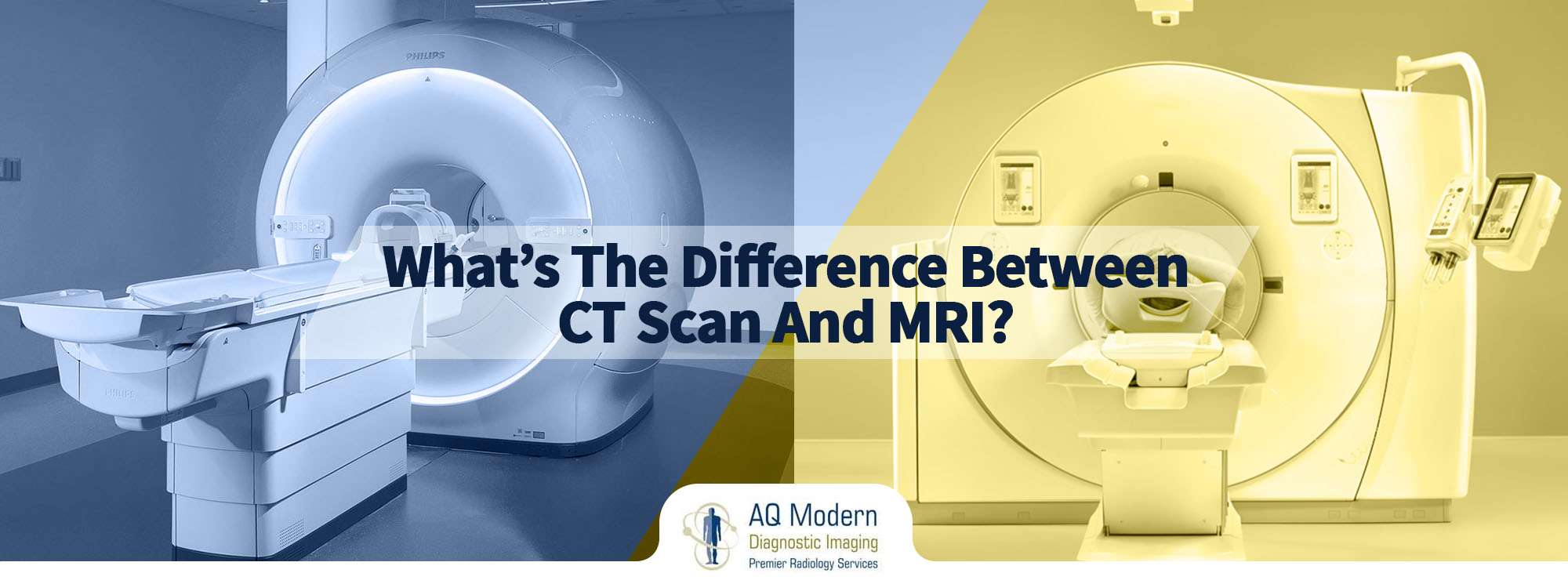
CT Machine vs MRI Machine: In relation to diagnostic imaging, there are two machines that stand out – Computed Tomography (CT) and Magnetic Resonance Imaging (MRI). Each machines are used to create detailed photographs of the within of the physique, however they work in essentially alternative ways and have totally different strengths and weaknesses.
On this article, we’ll delve into the world of CT and MRI machines, exploring their historic improvement, anatomy, working ideas, and scientific purposes. We’ll additionally focus on the elements that have an effect on picture decision and high quality, radiation publicity and security considerations, cost-effectiveness and accessibility, and technological developments and rising tendencies.
Variations between Computed Tomography (CT) Machines and Magnetic Resonance Imaging (MRI) Machines
Within the medical discipline, Computed Tomography (CT) machines and Magnetic Resonance Imaging (MRI) machines are two essential imaging modalities used to diagnose, deal with, and handle a variety of medical circumstances. Whereas each applied sciences play a significant position in fashionable drugs, every has distinct benefits, limitations, and purposes.
Historic Growth of CT and MRI Applied sciences
Computed Tomography (CT) know-how has its roots within the early Seventies, when Sir Godfrey Hounsfield developed the primary CT scanner. This pioneering work constructed upon earlier analysis in computed tomography, which relied on earlier analog computed tomography (CT) know-how first patented by Sir Godfrey Hounsfield’s predecessor. Sir Allan McLeod Cormack. The primary business CT scanner was launched in 1971, revolutionizing medical imaging by enabling high-resolution, cross-sectional photographs of the physique.
In distinction, Magnetic Resonance Imaging (MRI) know-how emerged within the Seventies and Nineteen Eighties, pushed by important advances in physics and engineering. The invention of nuclear magnetic resonance (NMR) within the Nineteen Forties laid the muse for MRI improvement. The primary MRI machines have been developed within the Seventies and early Nineteen Eighties, enabling the visualization of inside physique buildings utilizing magnetic fields and radio waves.
Rôle of CT and MRI Machines in Diagnostic Imaging
CT machines are broadly utilized in scientific settings for a variety of purposes, together with:
- Figuring out vascular illnesses, reminiscent of atherosclerosis and aneurysms.
- Detecting stomach and pelvic illnesses, reminiscent of appendicitis and kidney stones.
- Visualizing bone buildings and detecting fractures and osteoporosis.
MRI machines, alternatively, are significantly helpful in diagnosing neurological circumstances, reminiscent of:
- Sinuses and facial bone points
- Cardiovascular illnesses, together with valvular illness.
- Stomach and breast tissues points
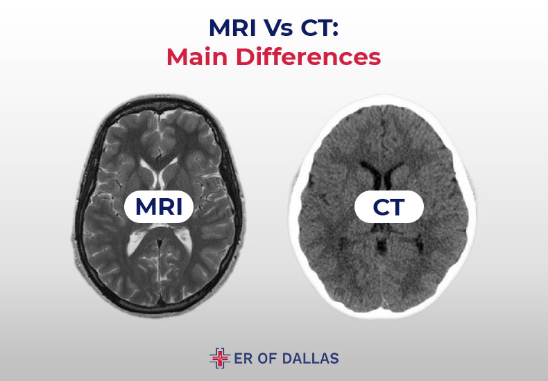
CT scans make the most of X-rays and laptop algorithms to reconstruct detailed photographs of physique buildings. CT scans are extremely efficient for detecting calcified buildings and lung illnesses. MRI machines use highly effective magnetic fields and radio waves to generate detailed photographs of sentimental tissues and organs, together with tumors and different abnormalities.
Key Technical Variations between CT and MRI Machines
The first variations between CT and MRI machines lie of their imaging ideas, technical specs, and scientific purposes.
- Imaging Precept: CT machines use X-rays to generate photographs, whereas MRIs make the most of magnetic fields and radio waves.
- Picture Decision: MRI machines usually supply increased spatial decision and distinction decision in comparison with CT scans.
- Medical Purposes: CT scans are sometimes used for emergency imaging, whereas MRI machines are extra generally employed for non-invasive prognosis and follow-up research.
The selection between CT and MRI machines usually is determined by the affected person’s particular situation, medical historical past, and the diagnostic necessities of the scientific state of affairs. Healthcare professionals fastidiously choose essentially the most appropriate imaging modality to acquire the very best diagnostic data.
Benefits of CT and MRI Machines
Each CT and MRI machines have distinct benefits that contribute to their widespread use in medical imaging.
- CT Machines: Fast picture acquisition, excessive accuracy for calcified buildings, and low price.
- MRI Machines: Excessive spatial decision, wonderful soft-tissue distinction, and the flexibility to visualise inside buildings with out ionizing radiation.
By recognizing the strengths and weaknesses of every imaging modality, healthcare professionals could make knowledgeable selections about diagnostic imaging, resulting in extra correct diagnoses and efficient affected person care.
Varieties of CT Machines and MRI Machines: Ct Machine Vs Mri Machine
CT (Computed Tomography) and MRI (Magnetic Resonance Imaging) machines are two of essentially the most broadly used medical imaging applied sciences in fashionable healthcare. Whereas each applied sciences have their very own distinct options and purposes, they share a standard aim: to supply diagnostic photographs of inside buildings and abnormalities throughout the human physique.
Varieties of CT Machines
There are a number of kinds of CT machines obtainable, every designed to cater to particular imaging wants and necessities. The principle kinds of CT machines embrace:
- Standard CT Scanners: These machines use a rotating X-ray supply and detector to supply cross-sectional photographs of the physique. They’re generally used for routine imaging purposes reminiscent of chest and stomach scans.
- Excessive-Pace CT Scanners: These machines use superior know-how to quickly purchase a number of slices of knowledge in a single rotation of the X-ray supply. They are perfect for purposes that require high-speed imaging, reminiscent of cardiac imaging.
- CT Scans with Distinction: The sort of CT machine makes use of a distinction agent to boost the distinction between totally different tissues and abnormalities throughout the physique. It’s generally used for imaging purposes that require detailed visualization of blood vessels and tumors.
Varieties of MRI Machines
MRI machines additionally are available in quite a lot of varieties, every designed to cater to particular imaging wants and necessities. The principle kinds of MRI machines embrace:
- Closed-Bore MRI Machines: These machines have a slender, enclosed bore that’s perfect for imaging purposes that require high-field power. They’re generally used for purposes reminiscent of mind and backbone imaging.
- Open-Bore MRI Machines: These machines have a wider, extra open bore that gives higher entry for sufferers with mobility points. They are perfect for purposes that require imaging of bigger physique components, such because the chest and stomach.
- Magnetic Resonance Spectroscopy (MRS) Machines: These machines use superior know-how to accumulate detailed spectroscopic knowledge from particular areas of the physique. They’re generally used for purposes that require detailed evaluation of biochemical processes throughout the physique.
Utility Areas for Every Kind of Machine
CT and MRI machines are broadly utilized in varied scientific and analysis purposes. Listed below are some frequent utility areas for every sort of machine:
- CT Machines:
- Routine imaging purposes reminiscent of chest and stomach scans.
- Cardiac imaging purposes that require high-speed imaging.
- Bariatric imaging purposes that require enhanced distinction agent capabilities.
- MRI Machines:
- Mind and backbone imaging purposes that require high-field power.
- Chest and stomach imaging purposes that require open-bore designs.
- Biochemical imaging purposes that require MRS capabilities.
Picture Decision and High quality

Picture decision and high quality are essential elements in medical imaging, enabling healthcare professionals to precisely diagnose and deal with varied medical circumstances. On this context, it’s important to know the elements that affect picture decision and high quality in each Computed Tomography (CT) and Magnetic Resonance Imaging (MRI) machines.
Picture Decision in CT Scans
In CT scans, a number of elements have an effect on the picture decision, together with the standard of the X-ray tube, the detector array, and beam collimation. The X-ray tube is accountable for producing X-rays that penetrate the physique and move by the detectors. The detector array, alternatively, consists of a number of sensors that seize the indicators from the X-rays. The beam collimation refers back to the adjustment of the X-ray beam to make sure that it’s targeted on the area of curiosity. A well-collimated beam ensures that the X-rays move by the identical space of the physique, leading to a transparent and detailed picture.
As an illustration, a high-quality X-ray tube with a excessive output capability can produce photographs with increased decision, whereas a well-designed detector array with a excessive pixel density can seize extra detailed data. Equally, correct beam collimation ensures that the picture is just not distorted by scattered radiation.
-
X-ray tube high quality
-
Detector array high quality
-
Beam collimation adjustment
These elements collectively contribute to the picture decision of CT scans, enabling healthcare professionals to diagnose and deal with varied medical circumstances successfully.
Picture High quality in MRI Machines
In MRI machines, the magnetic discipline power, receiver coils, and knowledge processing affect the picture high quality. The magnetic discipline power, usually measured in Tesla, determines the flexibility of the machine to differentiate between totally different tissues. Receiver coils, alternatively, seize the indicators emitted by the spinning nuclei, whereas knowledge processing performs a essential position in reconstructing the ultimate picture. A high-quality MRI machine with a powerful magnetic discipline and delicate receiver coils can produce photographs with excessive decision and element.
A better magnetic discipline power (usually 1.5 Tesla or 3 Tesla) allows the machine to differentiate between totally different tissues extra precisely, leading to higher picture high quality. Delicate receiver coils can seize extra detailed data, whereas superior knowledge processing algorithms can reconstruct photographs with increased decision and accuracy.
-
Magnetic discipline power
-
Receiver coil high quality
-
Information processing algorithms
These elements collectively contribute to the picture high quality of MRI machines, enabling healthcare professionals to diagnose and deal with varied medical circumstances successfully.
Examples of Greater Picture Decision Benefiting Healthcare
Greater picture decision might be helpful in varied medical eventualities, reminiscent of:
-
Diagnosing mind tumors and different neurological issues, the place correct imaging is essential for remedy planning.
-
Visualizing blood vessels and detecting vascular illnesses, the place high-resolution photographs may also help determine blockages and different abnormalities.
-
Diagnosing musculoskeletal issues, the place high-resolution photographs may also help determine tears, fractures, and different accidents.
These examples illustrate the significance of picture decision in medical imaging and spotlight the advantages of high-quality CT and MRI machines in healthcare.
Radiation Publicity and Security Issues
Radiation publicity is a essential side of Computed Tomography (CT) scans, with ionizing radiation posing potential dangers to sufferers. CT scans make use of X-rays to supply detailed cross-sectional photographs of the physique. The radiation dose required for a CT scan is considerably increased than that of typical X-rays. To place this into perspective, it’s estimated {that a} typical chest CT scan delivers a radiation dose roughly equal to 100-200 typical chest X-rays.
Radiation Publicity Dangers
Extended or repeated publicity to X-rays utilized in CT scans can improve the danger of most cancers, significantly in kids, girls of childbearing age, and anybody with a household historical past of most cancers. Nevertheless, the precise relationship between radiation publicity and most cancers threat remains to be a subject of ongoing analysis and debate. In line with the American Most cancers Society, the lifetime threat of most cancers from a CT scan is extraordinarily low, affecting an estimated 1-2 most cancers instances per 10,000 CT scans carried out.
Minimizing Radiation Publicity
Radiation publicity might be minimized by using a number of methods:
- Utilizing the bottom dose of radiation obligatory to acquire diagnostic photographs, often called dose optimization.
- Using superior CT scanner applied sciences, reminiscent of ultra-low dose protocols.
- Limiting the variety of CT scans, significantly in pediatric sufferers or these with a household historical past of most cancers.
- Utilizing various imaging modalities, like Magnetic Resonance Imaging (MRI) or Ultrasonography, when possible.
Working MRI Machines Security Precautions
Security precautions and protocols for working MRI machines embrace:
Magnetic Area Security Precautions, Ct machine vs mri machine
MRI machines make use of a powerful magnetic discipline to generate detailed photographs of the physique. Nevertheless, this magnetic discipline can pose potential dangers to sufferers and medical workers. To make sure secure operation, MRI machines are outfitted with a number of security options, together with:
- Static magnetic discipline limits, often within the vary of 0.1-3 Tesla, relying on the precise MRI machine mannequin.
- Steel screening of sufferers to determine potential ferromagnetic objects that could possibly be displaced or drawn to the magnetic discipline throughout scanning.
- Automated shutdown mechanisms in case of an emergency or tools malfunction.
Security Issues with Ferromagnetic Implants
Ferromagnetic implants, reminiscent of surgical clips or prosthetic joints, can pose a big threat throughout MRI procedures. In excessive instances, these objects can turn into displaced or drawn to the magnetic discipline, resulting in critical hurt or damage.
- A complete medical historical past must be taken earlier than an MRI process to determine any potential ferromagnetic implants.
- Sufferers with ferromagnetic implants could require particular MRI sequences or various imaging modalities.
- Medical workers must be educated in emergency response procedures, together with tools shutdown and evacuation protocols.
Claustrophobia and Anxiousness
Some people could expertise claustrophobia or nervousness throughout MRI procedures, significantly in open or closed-bore scanners. To handle these considerations, fashionable MRI machines usually make use of options reminiscent of:
- Open or wide-bore designs to scale back affected person confinement.
- Sedation or anesthesia to reduce nervousness.
- Quite a few distraction methods, reminiscent of soothing voices, music, or visible aids.
Medical Purposes and Diagnostics
Computed Tomography (CT) machines and Magnetic Resonance Imaging (MRI) machines play pivotal roles in fashionable medical diagnostics. Their purposes in emergency drugs, musculoskeletal imaging, and neurological diagnostics have tremendously enhanced the accuracy and pace of medical diagnoses.
Emergency Medication: Trauma Analysis and Acute Stroke
CT machines are extensively utilised in emergency settings for fast prognosis of acute circumstances, reminiscent of trauma and acute stroke. The scans present essential data on the extent of accidents or injury to the mind, enabling speedy intervention and saving treasured time. As an illustration, within the case of a affected person who has suffered a head damage, a CT scan may also help determine potential haemorrhages, swelling, or different problems that require pressing medical consideration. In acute stroke prognosis, CT scans help clinicians in detecting the presence of a blood clot, permitting them to find out the simplest course of remedy.
In acute stroke prognosis, there exists a big distinction between the roles of CT and MRI machines. CT scans can quickly detect haemorrhagic strokes, which account for roughly 10 p.c of all stroke instances, and allow medical professionals to make knowledgeable selections about remedy. However, MRI scans have a higher sensitivity in detecting ischaemic strokes and figuring out the affected areas of the mind.
Musculoskeletal Imaging and Neurological Diagnostics
MRI machines are broadly employed in musculoskeletal imaging for the prognosis of assorted circumstances affecting the musculoskeletal system. These circumstances embrace musculoskeletal tumours, degenerative joint illnesses, tendonitis, and ligament sprains. MRI scans present detailed photographs of sentimental tissues, reminiscent of tendons, ligaments, and cartilage, which are sometimes compromised in these circumstances.
Neurological diagnostics involving MRI scans have additionally reworked the best way clinicians diagnose and handle neurological issues. MRI scans can detect abnormalities within the mind and spinal twine, reminiscent of a number of sclerosis, spinal twine accidents, and mind tumours, facilitating focused remedy and bettering affected person outcomes. The flexibility of MRI know-how has led to its widespread adoption in neuroimaging, permitting for exact visualisation of mind anatomy and performance.
Comparability of CT and MRI Purposes
The selection between CT and MRI machines largely is determined by the precise situation being identified. CT scans are finest suited to detecting bone fractures, calcifications and sure vascular circumstances, whereas MRI scans excel in soft-tissue imaging and are extra correct in detecting neurological issues, tumours, and musculoskeletal accidents.
A essential issue to think about in CT versus MRI comparisons is their distinct properties, which allow them to visualise totally different points of the physique. This enables clinicians to tailor their diagnostic strategy to the precise wants of every affected person.
CT scans are sooner and extra handy, whereas MRI scans supply superior picture decision, enabling clinicians to diagnose and monitor advanced circumstances extra precisely. The final word aim of recent medical diagnostics is to realize exact, focused diagnoses, enabling the simplest administration and remedy of assorted medical circumstances.
Price-Effectiveness and Accessibility
Computed Tomography (CT) and Magnetic Resonance Imaging (MRI) machines are two important diagnostic instruments in fashionable healthcare. Whereas each machines present essential imaging knowledge, they’ve distinct price profiles and accessibility considerations.
The procurement and upkeep prices of CT and MRI machines are substantial, with MRI machines being considerably dearer to accumulate and keep because of the high-energy magnets and complicated tools concerned. In line with a examine by the Company for Healthcare Analysis and High quality (AHRQ), the common price of a brand new CT scanner is roughly $500,000, whereas an MRI machine can price upwards of $3 million. These prices have important implications for healthcare suppliers, significantly these with restricted budgets or in resource-constrained settings.
Relative Prices of CT and MRI Machines
The price-effectiveness of CT and MRI machines is a essential consideration for healthcare suppliers.
The prices related to CT and MRI machines might be damaged down into a number of parts, together with the preliminary buy worth, upkeep prices, and alternative components. These prices might be affected by varied elements, such because the machine’s age, utilization, and the producer.
Methods for Optimizing Useful resource Allocation and Prioritizing Sufferers for CT or MRI Imaging
Healthcare suppliers can make use of a number of methods to optimize useful resource allocation and prioritize sufferers for CT or MRI imaging based mostly on their medical wants. One strategy is to develop clear tips for when CT or MRI imaging is important, making an allowance for the affected person’s prognosis, medical historical past, and potential advantages of the imaging take a look at. One other technique is to encourage using various imaging modalities, reminiscent of ultrasound or X-ray, when doable.
Components Influencing Entry to CT and MRI Providers in Completely different Healthcare Settings
- Geographic Location: Healthcare services in rural or underserved areas could have restricted entry to CT and MRI machines because of logistical and monetary constraints.
- Monetary Sources: Healthcare suppliers with restricted budgets could wrestle to accumulate and keep CT and MRI machines, resulting in diminished entry to those providers.
- Availability of Skilled Employees: The correct operation and upkeep of CT and MRI machines require specialised coaching and experience. A scarcity of educated workers can compromise entry to those providers.
Technological Developments and Rising Tendencies
Computer systems Tomography (CT) and Magnetic Resonance Imaging (MRI) machines have been evolving at a fast tempo, providing improved picture high quality, diminished radiation publicity, and enhanced diagnostic capabilities. Researchers and producers are regularly engaged on upgrading these applied sciences to supply higher outcomes and extra comfy experiences for sufferers.
Twin-Vitality CT and Section-Distinction CT Developments
Analysis and improvement efforts have resulted within the creation of Twin-Vitality CT (DECT) and Section-Distinction CT (PCCT) methods. These applied sciences allow imaging of assorted supplies and substances throughout the physique with enhanced precision.
– Twin-Vitality CT makes use of two distinct power ranges to differentiate between totally different supplies, offering improved bone and lung imaging.
– Section-Distinction CT makes use of the properties of sunshine to seize detailed photographs of sentimental tissues, reminiscent of blood vessels and organs.
Superior MRI Applied sciences
The MRI know-how discipline has been seeing important developments, resulting in new prospects and insights in medical diagnostics. Practical MRI (fMRI) and Diffusion Tensor Imaging (DTI) are among the many key examples of this progress.
– Practical MRI detects modifications in mind exercise, permitting researchers to map neural connections and monitor illness development.
– Diffusion Tensor Imaging reconstructs tissue buildings, offering invaluable details about the mind’s white matter tracts and neural pathways.
Rising MRI Applied sciences
A number of cutting-edge MRI applied sciences are at the moment being explored. Ultrahigh-Area MRI, for example, is providing higher-resolution photographs, improved sensitivity, and higher diagnostic capabilities. One other improvement is Hybrid MRI, which mixes a number of imaging modalities to supply complete details about a affected person’s situation.
– Ultrahigh-Area MRI machines function at increased magnetic discipline strengths, enabling sharper photographs and extra correct diagnoses.
– Hybrid MRI methods mix MRI with different imaging methods, reminiscent of PET and CT scans, to supply a extra detailed understanding of illness circumstances and affected person responses to remedy.
Future Instructions and Potential Advantages
Developments in CT and MRI applied sciences are anticipated to result in improved diagnostic accuracy, diminished radiation publicity, and enhanced affected person experiences. Some potential purposes of those applied sciences embrace personalised drugs, illness monitoring, and remedy response evaluation.
– Personalised drugs might be achieved through the use of superior imaging methods to tailor remedy plans to particular person sufferers.
– Illness monitoring might be improved by monitoring modifications in illness development with the help of superior imaging applied sciences.
Closing Wrap-Up
As we have seen, CT and MRI machines are each highly effective instruments on the earth of diagnostic imaging, every with their very own strengths and weaknesses. Whereas CT machines are nice for visualizing bones, lungs, and different denser buildings, MRI machines excel at imaging delicate tissues, reminiscent of organs, tumors, and nerves. By understanding the variations between these two machines, we will higher recognize the significance of choosing the proper diagnostic imaging device for the job.
Whether or not you are a medical skilled or a curious affected person, understanding the fundamentals of CT and MRI machines may also help you navigate the world of diagnostic imaging with confidence.
Generally Requested Questions
What’s the distinction between a CT scan and an MRI scan?
A CT scan makes use of X-rays to create detailed cross-sectional photographs of the physique, whereas an MRI scan makes use of highly effective magnets and radio waves to create detailed photographs of sentimental tissues and organs.
Which imaging modality is healthier for diagnosing lung most cancers?
CT scans are usually higher than MRI scans for diagnosing lung most cancers, as they’re significantly efficient at detecting tumors and abnormalities within the lungs.
Can MRI machines detect blood clots within the mind?
Sure, MRI machines are extremely efficient at detecting blood clots within the mind, and are sometimes utilized in emergency conditions.
Are CT and MRI scans secure for pregnant girls?
Whereas each CT and MRI scans are usually secure for pregnant girls, it’s important to debate any considerations or dangers with a healthcare supplier earlier than present process both take a look at.

