`
`
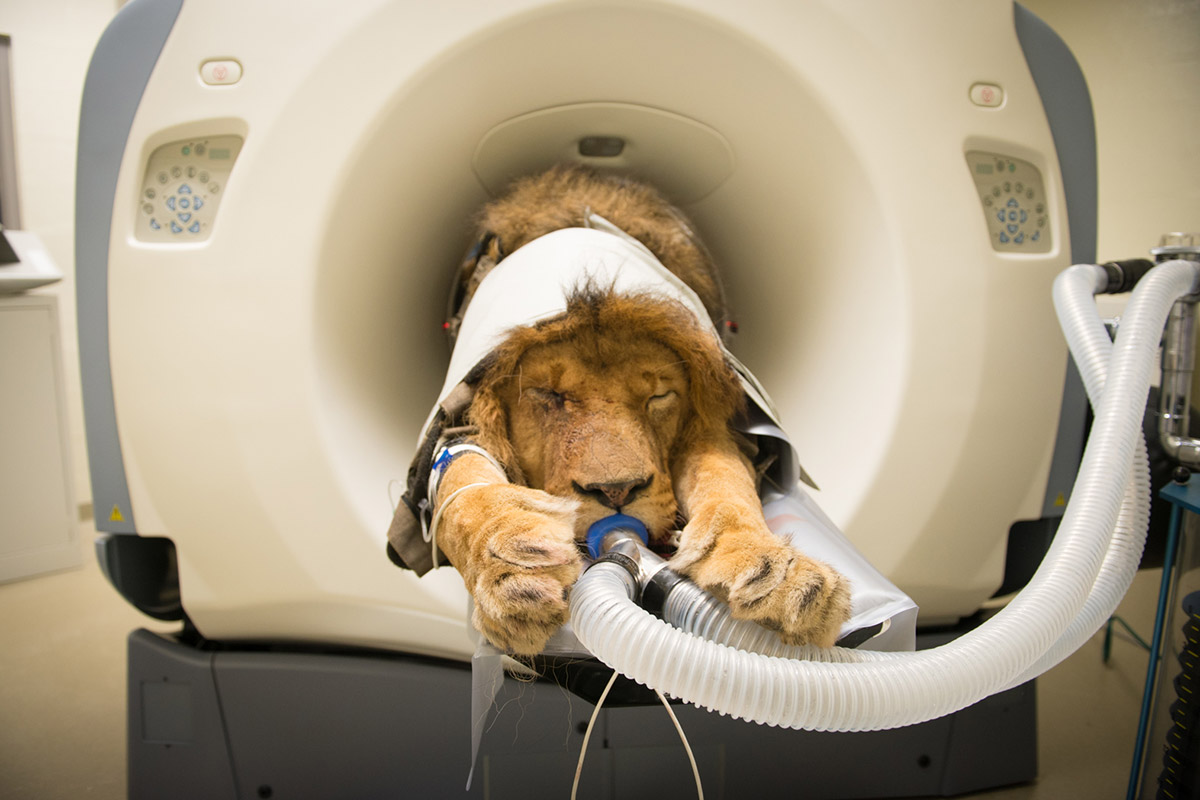
Cat Scan Machine Photographs contain a fancy course of of making detailed pictures of the physique’s inside buildings utilizing computed tomography (CT) scans. This expertise makes use of x-ray expertise to provide cross-sectional pictures that may be reconstructed into three-dimensional fashions for additional evaluation and analysis.
`
`
The CT scan course of includes a number of phases, together with affected person preparation, scanning, and picture reconstruction. Through the scanning course of, the affected person is positioned throughout the CT scanner, which makes use of x-ray beams to emit radiation by the physique. The detectors on the opposite aspect of the scanner report the attenuation of those x-rays as they go by totally different tissues, producing detailed pictures of the interior buildings.
`
`
Overview of Cat Scan Machine Photographs
Computed tomography (CT) scans are a non-invasive medical imaging method used to provide detailed cross-sectional pictures of the physique’s inside buildings. These pictures are important for diagnosing a variety of medical circumstances, together with accidents, tumors, and illnesses that have an effect on varied organs and tissues.
The imaging course of concerned in creating CT scan pictures relies on x-ray expertise. This is the way it works:
Position of X-ray Know-how in CT Scans
X-ray expertise is the cornerstone of CT scans. A CT scan makes use of a specialised x-ray machine to provide detailed pictures of the physique’s inside buildings. When a affected person undergoes a CT scan, they’re positioned contained in the scanner, which rotates round their physique, taking a number of x-ray pictures from totally different angles. These pictures are then reconstructed right into a two-dimensional illustration of the physique’s inside buildings.
The x-ray expertise utilized in CT scans is extra refined than conventional x-ray imaging. As an alternative of a single x-ray beam, a CT scanner makes use of a beam of x-rays that’s rotated across the physique, permitting for the creation of exact cross-sectional pictures.
Imaging Strategy of CT Scans
The imaging technique of CT scans includes the next steps:
- Positioning: The affected person is positioned contained in the scanner, and the scanner’s gantry (the ring that surrounds the affected person) is centered over the realm of curiosity.
- Knowledge Acquisition: The scanner takes a number of x-ray pictures from totally different angles because it rotates across the physique.
- Knowledge Reconstruction: The x-ray pictures are reconstructed right into a two-dimensional illustration of the physique’s inside buildings utilizing refined pc algorithms.
- Picture Show: The reconstructed pictures are displayed on a monitor, permitting medical doctors to visualise the interior buildings of the physique in nice element.
Through the use of x-ray expertise and superior pc algorithms, CT scans present medical doctors with a wealth of details about the physique’s inside buildings, enabling correct diagnoses and efficient remedy of a variety of medical circumstances.
Objective of Computed Tomography (CT) Scans
Computed tomography (CT) scans are used to diagnose a variety of medical circumstances, together with accidents, tumors, and illnesses that have an effect on varied organs and tissues. The aim of a CT scan is to offer medical doctors with detailed pictures of the physique’s inside buildings, permitting for correct diagnoses and efficient remedy.
A few of the widespread medical circumstances identified utilizing CT scans embrace:
- Bone fractures and accidents
- Tumors and cancers
- Liver illness and cirrhosis
- Kidney illness and kidney stones
- Pneumonia and different lung circumstances
CT scans are additionally used to information interventional procedures, resembling biopsies and tumor biopsies, and to observe the development of illnesses over time.
Picture Acquisition Methods
The method of scanning a affected person with a CT scanner includes a number of steps, beginning with the preparation of the affected person and ending with the reconstruction of the picture. The affected person is often positioned contained in the scanner, which is a donut-shaped machine that rotates across the affected person to seize a number of cross-sectional pictures.
Scanning Course of
The scanning course of is often initiated by the technologist who operates the CT scanner. The technologist units the suitable scanning parameters, such because the scan slice thickness, reconstruction algorithm, and discipline of view. The affected person is then positioned contained in the scanner, and the scanner begins to rotate across the affected person, emitting X-rays to penetrate the affected person’s physique. The X-rays are then detected by a number of sensors, which ship the information to the pc for reconstruction.
The scanning course of sometimes takes a number of minutes, and the affected person is requested to stay nonetheless throughout this time to keep away from movement artifacts within the picture.
Picture Decision and Scanning Parameters
The picture decision of a CT scan is affected by a number of scanning parameters, together with the slice thickness, reconstruction algorithm, and radiation dose. The slice thickness refers back to the thickness of every particular person picture slice, and it will probably vary from just a few millimeters to a number of centimeters. The reconstruction algorithm is the software program used to reconstruct the picture from the uncooked information, and it will probably have an effect on the picture high quality and determination.
The radiation dose, also called the publicity time, can even have an effect on the picture decision. The next radiation dose can present larger picture decision, however it will probably additionally improve the affected person’s publicity to radiation.
Significance of Distinction Brokers
Distinction brokers are substances which are injected into the affected person’s blood vessels to reinforce the visibility of sure buildings or lesions within the picture. Distinction brokers are notably helpful in vascular imaging, resembling coronary artery illness or stroke. They work by absorbing X-rays and permitting the scanner to detect them, making the buildings or lesions seem brighter within the picture.
The most typical forms of distinction brokers utilized in CT scans are iodine-based brokers, that are utilized in vascular imaging, and barium-based brokers, that are utilized in gastrointestinal imaging.
- Distinction brokers can be utilized to reinforce the visibility of sure buildings or lesions within the picture, making it simpler to diagnose and deal with illnesses.
- The selection of distinction agent is dependent upon the kind of imaging being carried out and the affected person’s medical historical past.
- Distinction brokers could cause allergic reactions, so sufferers are sometimes requested to offer a medical historical past earlier than present process a CT scan with distinction.
Picture Reconstruction Algorithms

Picture reconstruction algorithms play a vital function inComputed Tomography (CT) scans by remodeling the uncooked information acquired from the CT scanner into detailed pictures of the interior buildings of the physique. Varied algorithms are used to reconstruct pictures, every with its personal strengths and weaknesses. This part will discover the ideas behind filtered backprojection (FBP) reconstruction, some great benefits of iterative reconstruction algorithms, and the function of beam hardening correction in CT scan picture reconstruction.
Filtered Backprojection (FBP) Reconstruction
Filtered backprojection is among the earliest and most generally used algorithms for picture reconstruction in CT scans. The precept behind FBP reconstruction includes making use of a filter to the projection information to scale back noise and artifacts, after which backprojecting the filtered information to reconstruct the picture.
The filtered backprojection algorithm may be mathematically represented as:
the place R(x,y) is the reconstructed picture, P(x,y,z) is the projection information, and H(x,y,z) is the filter operate.
Filtered backprojection is broadly utilized in CT scans as a consequence of its simplicity and pace. Nevertheless, it has some limitations, together with:
* Noise sensitivity
* Restricted skill to get well small particulars
* Over-smoothing of huge buildings
Iterative Reconstruction Algorithms
Iterative reconstruction algorithms are extra superior and have been gaining reputation in recent times as a consequence of their superior picture high quality and elevated robustness to noise. These algorithms reconstruct the picture by iteratively updating the picture estimate primarily based on the projection information and a set of regularization equations.
Iterative reconstruction algorithms have a number of benefits over FBP reconstruction, together with:
* Improved picture high quality
* Elevated robustness to noise
* Means to get well small particulars
* Higher dealing with of high-contrast objects
The most typical iterative reconstruction algorithm is the Most Chance Expectation Maximization (MLEM) algorithm, which may be mathematically represented as:
<βk+1 = βk + γ * ∇L(βk|P)>
the place βk is the picture estimate at iteration ok, γ is the step dimension, L(βk|P) is the probability operate, and ∇L(βk|P) is the gradient of the probability operate.
Beam Hardening Correction
Beam hardening correction is a crucial step in CT scan picture reconstruction, notably for high-contrast objects. It includes correcting for the attenuation of the X-ray beam because it passes by the article, which may result in artifacts and inaccuracies within the picture reconstruction.
Beam hardening may be mathematically represented as:
the place I(x,y) is the reconstructed picture, ρ(x,y,z) is the density of the article, μ(E,z) is the attenuation coefficient, and E is the X-ray vitality.
Correcting for beam hardening includes making use of a correction issue to the projection information, which may be calculated utilizing varied strategies, together with:
* Polynomial becoming
* Nonlinear becoming
* Machine studying algorithms
Benefits of Iterative Reconstruction Algorithms over FBP
Iterative reconstruction algorithms have a number of benefits over FBP reconstruction, together with:
* Improved picture high quality
* Elevated robustness to noise
* Means to get well small particulars
* Higher dealing with of high-contrast objects
Some great benefits of iterative reconstruction algorithms may be summarized as follows:
* Improved distinction and determination
* Decreased noise and artifacts
* Elevated sensitivity to small lesions
* Higher dealing with of high-contrast objects
Conclusion
In conclusion, picture reconstruction algorithms play a vital function in CT scans by remodeling the uncooked information into detailed pictures of the interior buildings of the physique. FBP reconstruction is broadly used as a consequence of its simplicity and pace, however iterative reconstruction algorithms provide superior picture high quality and elevated robustness to noise. Beam hardening correction is a crucial step in CT scan picture reconstruction, notably for high-contrast objects.
Picture Processing and Submit-processing
Picture processing and post-processing are essential steps in enhancing the standard of CT scan pictures. These steps assist to take away noise, enhance distinction, and visualize the interior buildings of the physique. The aim of picture processing and post-processing is to provide high-quality pictures that help in correct analysis and remedy planning.
Enhancing CT Scan Photographs
Enhancing CT scan pictures includes adjusting the brightness and distinction to optimize the visualization of inside buildings. This may be achieved by varied methods, together with:
- Windowing: Windowing is a method used to regulate the distinction and brightness of CT scan pictures. It includes adjusting the window stage and width to optimize the visualization of particular buildings.
- Gamma correction: Gamma correction is a method used to regulate the brightness and distinction of CT scan pictures. It includes adjusting the gamma worth to optimize the visualization of particular buildings.
- Histogram equalization: Histogram equalization is a method used to regulate the distinction of CT scan pictures. It includes adjusting the histogram to optimize the visualization of particular buildings.
Gamma worth and histogram equalization play a vital function in enhancing pictures, as they regulate the brightness ranges and unfold of the photographs.
Decreasing Picture Noise in CT Scans
Picture noise in CT scans is brought on by varied elements, together with the X-ray beam, the detector, and the picture reconstruction algorithm. To scale back picture noise, varied methods may be employed, together with:
- Noise discount filters: Noise discount filters are algorithms which are used to scale back noise in CT scan pictures. They contain making use of a filter to the picture to take away noise and enhance picture high quality.
- Denoising algorithms: Denoising algorithms are algorithms which are used to take away noise from CT scan pictures. They contain making use of a mathematical operate to the picture to take away noise and enhance picture high quality.
- Photon counting: Photon counting is a method used to scale back noise in CT scans. It includes measuring the variety of photons which are detected by the detector to enhance picture high quality.
These methods might help scale back the noise ranges in CT scan pictures, resulting in improved picture high quality and accuracy.
The Position of Segmentation in CT Scan Picture Processing
Segmentation is an important step in CT scan picture processing that includes dividing the picture into totally different areas or segments. This may be achieved by varied methods, together with:
- Thresholding: Thresholding is a method used to section CT scan pictures. It includes making use of a threshold worth to the picture to separate the foreground from the background.
- Edge detection: Edge detection is a method used to section CT scan pictures. It includes detecting the sides of buildings within the picture to separate the foreground from the background.
- Machine studying algorithms: Machine studying algorithms are used to section CT scan pictures. They contain coaching a mannequin on a dataset of pictures to be taught the options of various buildings and section the picture accordingly.
Segmentation performs a vital function in CT scan picture processing, because it permits the visualization of particular buildings and helps in correct analysis and remedy planning.
Picture Registration
Picture registration is a necessary step in picture evaluation, which includes aligning two or extra pictures taken at totally different occasions or from totally different imaging modalities. This may be achieved by varied methods, together with:
- Function-based registration: Function-based registration includes registering pictures primarily based on widespread options resembling edges, strains, or factors.
- Depth-based registration: Depth-based registration includes registering pictures primarily based on their depth values.
- Mutual information-based registration: Mutual information-based registration includes registering pictures primarily based on the mutual data between the photographs.
Picture registration permits the comparability of pictures taken at totally different occasions or from totally different imaging modalities, which is crucial for assessing illness development, remedy response, and monitoring the effectiveness of therapies.
Picture processing and post-processing are important steps in CT scan imaging, which contain enhancing picture high quality and decreasing noise ranges. These steps are essential for correct analysis and remedy planning.
Purposes of CT Scan Imaging
CT scan imaging has quite a few functions within the medical discipline, revolutionizing the way in which varied illnesses are identified and handled. One of the vital important makes use of of CT scans is within the discipline of oncology.
Diagnosing Most cancers
CT scans play an important function in diagnosing most cancers by offering detailed pictures of inside organs and tissues. They assist medical doctors visualize the most cancers’s location, dimension, and extent, enabling them to develop an efficient remedy plan. CT scans can detect most cancers in varied phases, from early tumors to superior metastatic illness. They’ll additionally help in differentiating between benign and malignant tumors, serving to medical doctors make knowledgeable choices about the perfect course of remedy.
Position in Emergency Medication
In emergency drugs, CT scans are used to rapidly and precisely diagnose and deal with important circumstances resembling trauma, stroke, and inside bleeding. They supply medical doctors with very important details about the severity of accidents and the perfect remedy choices. CT scans are notably helpful in assessing sufferers with head trauma, as they will rapidly establish hemorrhages and different life-threatening circumstances. In stroke sufferers, CT scans might help medical doctors decide the kind of stroke and the affected areas of the mind, enabling them to provoke well timed remedy.
Orthopedic Imaging
CT scans are additionally broadly utilized in orthopedic imaging to diagnose and deal with joint issues. They supply detailed pictures of bones, joints, and surrounding tissues, enabling medical doctors to visualise fractures, degenerative modifications, and different circumstances. CT scans might help diagnose circumstances resembling osteoarthritis, rheumatoid arthritis, and tendonitis, permitting medical doctors to develop efficient remedy plans. They’ll additionally help in planning surgical procedures, resembling joint replacements and bone grafts.
Future Developments in CT Scan Know-how

The way forward for CT scan expertise is marked by vital developments and ongoing analysis. These developments intention to enhance the accuracy, pace, and security of CT scans, in addition to improve the general affected person expertise. On this part, we discover among the rising traits and improvements in CT scan expertise.
Synthetic Intelligence and Machine Studying in CT Scans
The combination of synthetic intelligence (AI) and machine studying (ML) into CT scan expertise has the potential to revolutionize the sphere. AI-powered CT scans can analyze pictures extra rapidly and precisely than human radiologists, decreasing the time it takes to diagnose and deal with sufferers. ML algorithms may be skilled to establish particular patterns and abnormalities in pictures, permitting for extra exact analysis and remedy. This expertise can even assist scale back the dose of radiation used throughout CT scans, making them safer for sufferers.
Potential Advantages of Hybrid CT-PET Scanners, Cat scan machine pictures
Hybrid CT-PET scanners mix the capabilities of each computed tomography (CT) and positron emission tomography (PET) to offer a extra complete understanding of the physique’s inside buildings and capabilities. This expertise permits medical doctors to see each the morphology (form and construction) and performance (metabolic exercise) of tissues and organs in a single scan. This may result in extra correct diagnoses, diminished remedy occasions, and improved affected person outcomes.
Developments in Computed Tomography Angiography (CTA)
Computed tomography angiography (CTA) is a specialised imaging method that makes use of CT scans to visualise the blood vessels and establish any potential blockages or abnormalities. Current developments in CTA expertise have led to improved picture decision, quicker scan occasions, and diminished radiation doses. These developments have made CTA a priceless software for diagnosing and treating cardiovascular illnesses, resembling atherosclerosis and stroke.
Rising Traits and Future Instructions
Extra rising traits and future instructions in CT scan expertise embrace:
- Quantitative imaging, which makes use of mathematical calculations to investigate and quantify imaging information.
- Picture-guided procedures, which use real-time imaging to information biopsies, tumor removals, and different minimally invasive procedures.
- Spectral CT imaging, which makes use of a number of vitality settings to provide detailed pictures of the physique’s inside buildings and capabilities.
Closing Abstract
`
`
General, the evaluation of cat scan machine pictures is a important step within the analysis and remedy of varied medical circumstances. By understanding the ideas of CT scan expertise and picture reconstruction, healthcare professionals can produce high-quality pictures that inform medical choices and enhance affected person outcomes.
`
`
Future developments in CT scan expertise, together with developments in synthetic intelligence and hybrid CT-PET scanners, maintain promise for much more correct and detailed imaging. As this expertise continues to evolve, it’s important to keep up a concentrate on affected person security and radiation publicity.
`
`
Frequent Queries
`
`
What’s the main function of a CT scan?
`
`
A CT scan is used to provide detailed cross-sectional pictures of the physique’s inside buildings utilizing x-ray expertise.
`
`
How does a CT scanner work?
`
`
A CT scanner makes use of x-ray beams to emit radiation by the physique, which is recorded by detectors on the opposite aspect of the scanner. These recordings are then used to provide detailed pictures of the interior buildings.
`
`
What’s the function of distinction brokers in CT scans?
`
`
Distinction brokers are used to reinforce the visibility of particular tissues or buildings throughout the physique by altering the attenuation of x-rays.
`
`
What’s the distinction between axial, coronal, and sagittal CT scan pictures?
`
`
These pictures are obtained from totally different views of the physique, with axial pictures obtained from the back and front, coronal pictures obtained from the aspect, and sagittal pictures obtained from the highest and backside.
`
`
How do I put together for a CT scan?
`
`
Preparation sometimes includes eradicating any steel objects, carrying unfastened clothes, and probably receiving a distinction agent injection. The particular preparation steps will range relying on the scan sort and medical situation being evaluated.
`
`

